




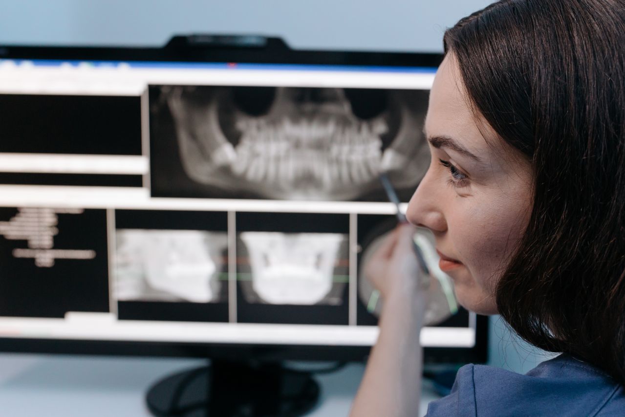
You might be sitting in the dental chair, bib clipped on, when the assistant at a practice focused on family dentistry in Wichita Falls pulls out the X ray sensor. A quiet thought pops up. “Is this safe? How much radiation am I getting? Do I really need this?” You are not being dramatic. You are being careful, and that is a good thing.end
Over the years, many people have heard conflicting things about dental X rays. Some are told they are totally harmless. Others read warnings online and start to worry about every image. It can feel like you are stuck between wanting clear answers and not wanting to sound anxious or “difficult.”
Here is the short version of what you need to know. Modern digital dental X rays are designed with your safety in mind. They use far less radiation than older film systems, they give your dentist clearer images to catch problems early, and they help avoid unnecessary repeat exposures. When used properly and only when needed, they are a powerful tool to protect your health, not a hidden threat to it.
So where does that leave you? You deserve to understand what is happening and why, so you can say yes to X rays with confidence instead of with a knot in your stomach.
Why do dental X rays feel scary, and what is really at stake?
Part of the fear comes from the word “radiation.” It sounds heavy and dangerous. You might picture hospital scanners, warning labels, or even headlines about overexposure. When you care about your health or your child’s health, those images stick.
On the other side, you know your teeth do not always tell the truth on the surface. Cavities can form between teeth where you cannot see them. Infections can start at the root of a tooth with almost no pain at first. Bone loss can quietly progress for years. Your dentist needs a way to see what your eyes cannot.
This tension creates a natural worry. “Am I trading long term risk for short term information?” It helps to know that organizations like the American Dental Association have clear guidance on when dental X rays are appropriate and how to keep exposure as low as reasonably achievable. You can read more about that in the ADA’s overview of dental X rays and radiographs.
So the real question becomes not “Are X rays good or bad?” but “How do newer digital dental radiographs change the safety picture compared with older methods?”
How do digital X rays reduce radiation exposure for dental patients?
Traditional film X rays needed more radiation to create a clear image. The film itself was less sensitive, and you had to “get it right” in one shot, because fixing a bad film often meant taking another image.
Digital sensors are much more sensitive to X ray energy. That means your dentist can use a dramatically lower dose while still getting a sharp, useful picture. In many cases, digital systems can cut exposure compared with old film by a large margin, while also allowing tighter control of the settings for each patient.
The U.S. Food and Drug Administration recognizes the importance of limiting exposure. They provide detailed advice on how dental teams should select patients for X ray exams and how often images are truly needed. If you want to see the framework your dentist is encouraged to follow, you can look at the FDA guidance on selecting patients for dental radiographic examinations.
Because digital systems are faster and more forgiving, your chance of a “retake” is lower. Fewer retakes mean fewer total exposures across your lifetime, which is exactly what you want.
Can better images from digital X rays actually keep you safer?
It might sound strange, but a higher quality X ray can be a safety feature. When your dentist can see small problems clearly, they can treat them when they are still simple and less invasive. That protects you from bigger procedures later.
Imagine a cavity starting between two back teeth. You may not feel anything. It might not show on the surface. A clear digital bitewing image can reveal that tiny shadow early. A small filling now is easier on your body, your schedule, and your wallet than a root canal or extraction years later.
For more complex cases, some dentists use 3D imaging, called cone beam computed tomography or CBCT. This is not for routine checkups. It is reserved for situations where 2D X rays are not enough, such as planning an implant or evaluating complicated root anatomy. The FDA has specific information on dental cone beam CT, because even advanced tools must be used thoughtfully and only when clearly helpful.
When your dentist chooses the right type of X ray for the right reason, those images become a safety net, catching silent problems before they turn into emergencies.
How do digital X rays support safer long term care and fewer surprises?
Safety is not just about a single exposure. It is about your whole story over many years. Digital systems make that long view easier.
Electronic images are stored in your record and can be compared side by side over time. Your dentist can track changes in bone level, monitor a suspicious area, or confirm that a root canal is healing. This reduces guesswork and helps avoid unnecessary treatments.
Because images can be shared securely with specialists, you also avoid repeat X rays when you see another provider. That means less radiation and less hassle for you. Your care team can collaborate using the same clear images, which improves safety during complex treatment like implants, orthodontics, or oral surgery.
So instead of thinking of X rays as a one time risk, you can start to see digital dental imaging as part of a broader safety strategy that gives your dentist a full, accurate picture while keeping exposure as low as possible.
Digital vs traditional dental X rays: what should you weigh?
When you are deciding how comfortable you feel with X rays, it can help to compare the older and newer approaches side by side. This is not about technical details. It is about the parts that matter to you as a patient.
| Aspect | Traditional Film X Rays | Digital Dental X Rays |
|---|---|---|
| Typical radiation dose | Higher, film is less sensitive | Lower, sensors are more sensitive and adjustable |
| Image quality | Good, but harder to adjust after exposure | High quality, can enhance contrast and zoom without retakes |
| Retake risk | More common if film is over or under exposed | Less common, preview is instant and settings can be fine tuned |
| Time to view | Requires chemical processing, more waiting | Appears almost instantly on screen |
| Record keeping | Physical films can be lost or damaged | Stored electronically, easy long term comparison |
| Environmental impact | Uses chemicals and physical film | No processing chemicals, less physical waste |
The point is not that film X rays are automatically unsafe. When used correctly, they have helped many people for many years. The point is that digital systems give your dental team more options to lower exposure, reduce repeat images, and care for you more precisely.
What can you do right now to make dental X rays safer for you?
You are not a passive passenger in this process. There are simple, respectful steps you can take so X rays feel like a shared decision instead of something that just happens to you.
1. Ask why each X ray is needed today
Before the sensor goes in, you can calmly ask, “Can you tell me what you are looking for with this X ray?” A thoughtful dental team will explain whether it is for routine cavity checks, to monitor a known problem, or to plan a specific treatment. This keeps your care aligned with guidelines, not just habit.
2. Share your health history and recent imaging
Tell your dentist if you have had many X rays or scans in other settings recently. Also mention if you are pregnant, trying to conceive, or managing a medical condition. Bring recent dental images if you have them. This helps your dentist avoid unnecessary repeats and tailor their recommendations to your situation.
3. Request protective measures and digital options
You can ask if the office uses digital X rays and how they limit exposure. It is reasonable to request a lead apron and thyroid collar if appropriate. You can say, “I want to keep my exposure as low as possible. Are these digital X rays, and how often do you usually take them in a case like mine?” A good team will not be annoyed. They will be glad you care.
Moving forward with more confidence and less fear
You do not have to choose between being safe and being informed. With modern digital dental X ray technology, the two go hand in hand. The radiation levels are lower than older systems, the images are clearer, and the ability to track and share those images makes your long term care more predictable and less risky.
If you have been putting off a checkup because you are uneasy about X rays, know that your concerns are valid and that you are allowed to ask questions. A good general dentist will welcome that conversation, walk you through the reasons for each image, and use the least amount of radiation needed to protect your oral health.
You deserve care that feels safe, respectful, and transparent. With the right information and a dental team that listens, digital X rays can become something you agree to with a steady breath, not something you fear in silence.